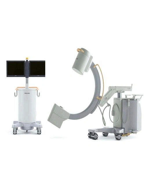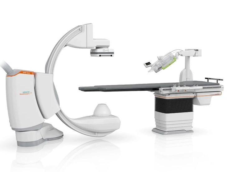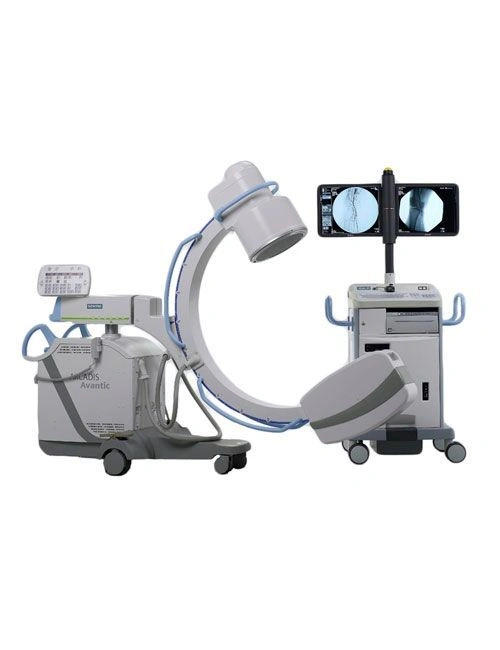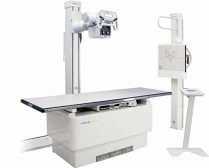
X-ray machines are advanced diagnostic devices that utilize high-energy electromagnetic radiation to generate images of the internal structures of objects and living organisms. Typically employed in medical environments, these machines are critical for diagnosing a variety of health conditions and monitoring patient responses to treatments. They consist of an X-ray tube that emits X-rays, a detector or film that captures these rays, and a control panel for settings adjustment. X-ray machines are categorized into three primary types: Radiography for still images, Fluoroscopy for real-time imaging, and Computed Tomography (CT) for cross-sectional views. The applications of X-ray machines span across various fields including medicine, dentistry, and industrial inspections. They typically carry safety features to ensure minimal risk of radiation exposure, utilizing protective equipment and technology advancements to enhance imaging quality while reducing radiation doses.
Key Features
| Features | Description |
|---|---|
| X-ray Type | Radiography, Fluoroscopy, Computed Tomography (CT) |
| Applications | Medical diagnosis, dental examinations, industrial inspection |
| Image Capture | Digital detectors for immediate display or traditional film |
| Control Options | Adjustable exposure time and radiation dose via control panel |
| Safety Features | Lead aprons, protective barriers, limited exposure time |
| Attributes | Description |
|---|---|
| X-ray Tube | High-performance tube for optimized X-ray emission |
| Image Quality | High-resolution images with advanced digital technology |
| Radiation Safety | Equipped with multiple safety mechanisms to minimize exposure |
| Diagnostic Capabilities | Effective for diagnosing fractures, infections, and tumors |
| Usability | User-friendly interface with control settings for ease of operation |
Key Words
*Disclaimer: The above description has been AI-generated and has not been audited or verified for accuracy. It is recommended to verify product details independently before making any purchasing decisions.
MSME Certificate
Fssai certificate
Incorporation Type
partnership firm
GST
06AARFN1109Q1ZT
Number of Employees
1 To 10 People
Import Export Code (IEC)
AUYPNXXXXX
Year of Establishment
January 2017
Nature of Business
retailer / manufacturer / service provider / service center / manufacturer / professional services / contractor / retailer / brand owner / distributor / exporter / importer
GlobalLinker Member Since
April 2023
Annual Turnover: 1.00 crore-5.00 crore
Industry: Agriculture & Agri Products > Agri Produce & Finished Products
Location(s): West Delhi (India) | Delhi (India)
NIRVAN ENTERPRISES Manufacturer, Exporter, Importer. Add : 457,4th Floor,Sector-19,Dwarka, New Delhi- 110075 M: +91-859554140,+91-9999180823 E-mail: ne@nirvans.co
Country Of Origin: India
X-ray machines are devices that use X-rays—high-energy electromagnetic radiation—to create images of the inside of objects, commonly used in medical settings to diagnose and monitor various conditions. The core components include an X-ray tube, which produces X-rays, and a detector or film that captures the images.
Radiography: Produces static images for diagnostics.
Fluoroscopy: Provides real-time imaging for procedures.
Computed Tomography (CT): Combines multiple X-ray images to create cross-sectional views.
Applications:
Medical: Diagnosing fractures, infections, tumors, and monitoring treatments.
Dental: Assessing dental health and planning treatments.
Industrial: Inspecting welds and structural integrity.
Safety: While X-ray exposure is generally low, measures are taken to minimize risk, including shielding, limiting exposure time, and using protective equipment.
Technological Advances: Developments include digital X-ray systems, which enhance image quality and reduce radiation exposure.
Overall, X-ray machines are invaluable tools in healthcare and industry, providing critical insights while prioritizing patient safety.
Detector/Film: Positioned opposite the X-ray tube, this captures the X-rays that pass through the body. Traditional systems use film, while modern machines often use digital detectors for immediate image display.
Control Panel: This allows operators to adjust settings like exposure time and radiation dose.
Functioning:
When X-rays are emitted from the tube, they penetrate the body or object. Different tissues absorb X-rays at varying rates: bones absorb more and appear white on the image, while softer tissues appear darker. This contrast helps in diagnosing conditions like fractures, tumors, and infections.
Types:
Radiography: Produces still images for diagnostics.
Fluoroscopy: Offers real-time video imaging for procedures like swallowing studies.
CT Scans: Create detailed cross-sectional images by taking multiple X-ray images from different angles.
Applications:
Medical: Widely used in hospitals and clinics for diagnostic purposes.
Dental: Essential for examining teeth and jaw structures.
Industrial: Employed in non-destructive testing to check the integrity of materials and structures.
Safety Considerations:
X-ray machines are designed with safety features to minimize radiation exposure. This includes using lead aprons, limiting exposure time, and employing protective barriers. Advances in technology have also led to reduced radiation doses while maintaining image quality.
Overall, X-ray machines play a crucial role in medicine and industry, providing essential insights into health and structural integrity.


Delhi, India
Service Provider , Service Center, Manufacturer, Professional Services, Contractor, Retailer, Brand Owner, Distributor, Exporter, Importer, Wholesaler, Women Entrepreneur, Startup
GST- 07AUYPN5229L1ZR
FIEO Registered